
Sooty Bark Disease
What is SBD | Identifying SBD | Sample Submission | Look-a-likes | Collaboration | Human Health | Additional Resources | Contact Us
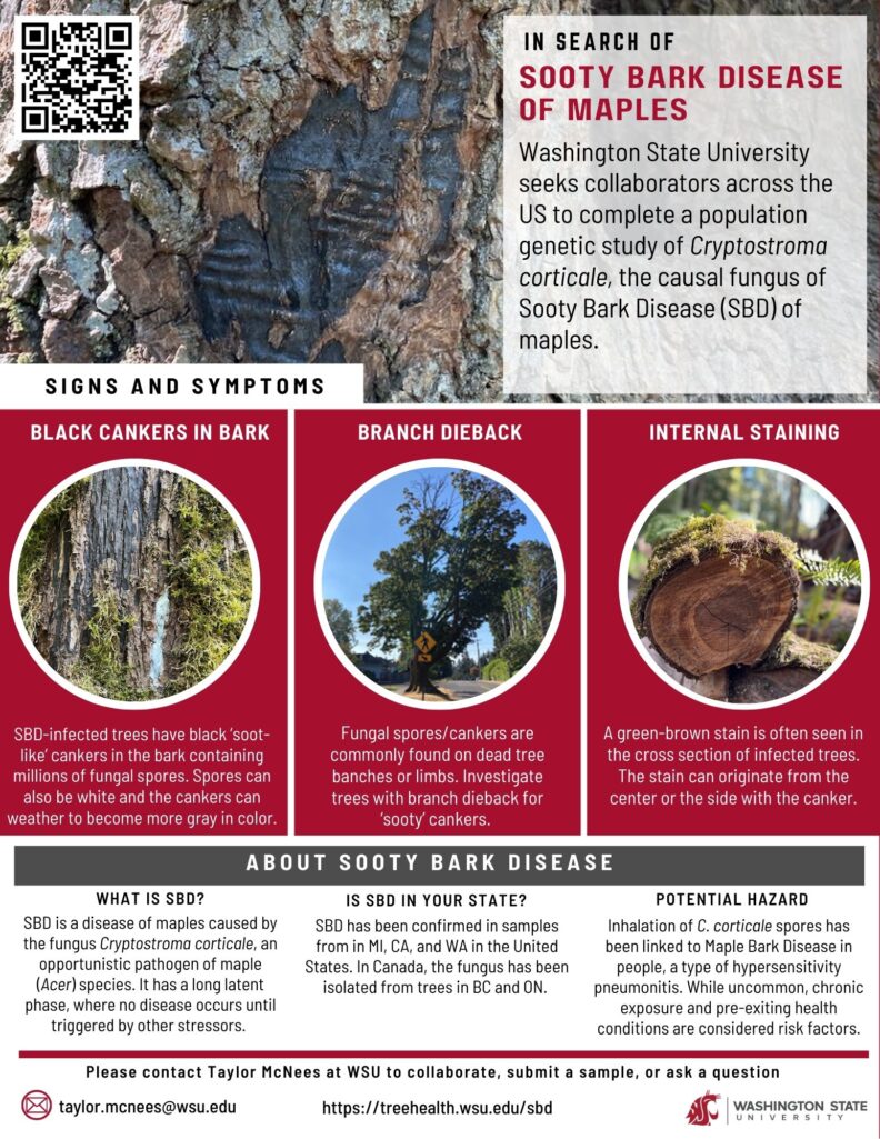
What is Sooty Bark Disease (SBD)?
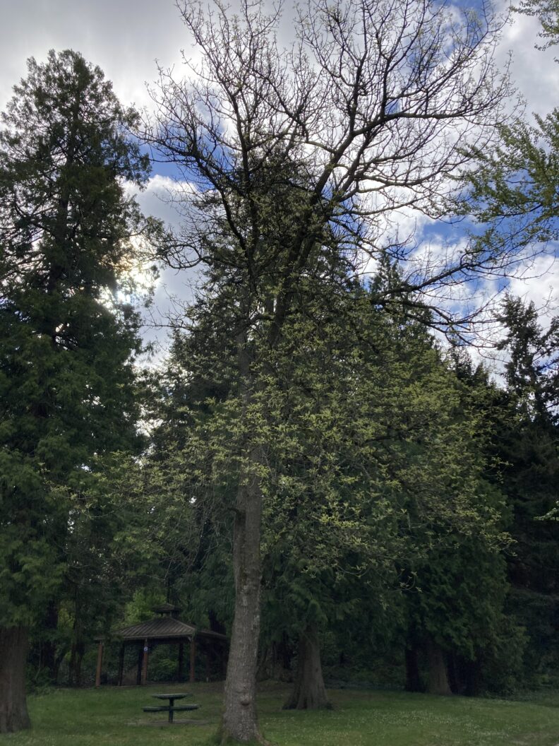
Sooty Bark Disease is an emerging concern within the Pacific Northwest. The disease, caused by the fungus Cryptostroma corticale, is known to primarily affect maple (Acer) species. The fungus forms black lesions within the bark of the infected stem/branch, where large numbers of spores begin to accumulate. Eventually, the buildup of spores causes the bark to shed and exposes the spores to be spread to new hosts by the wind.
It is hypothesized that C. corticale is an opportunistic pathogen that lives within its hosts for long periods of time without causing disease. Only once the host is exposed to other stress factors, such as heat or drought, does the switch to pathogenicity occur. Thus, it is predicted that with the changing climate and anticipated increase in extreme weather events will cause SBD to rise.
It has been well established that C. corticale infects Sycamore maples in Europe. However, recently the fungus has been identified on other species of maple, as well as some other tree species! However, to date the pathogenicity of the fungus has only been confirmed on sycamore and silver maple.
Identifying Sooty Bark Disease
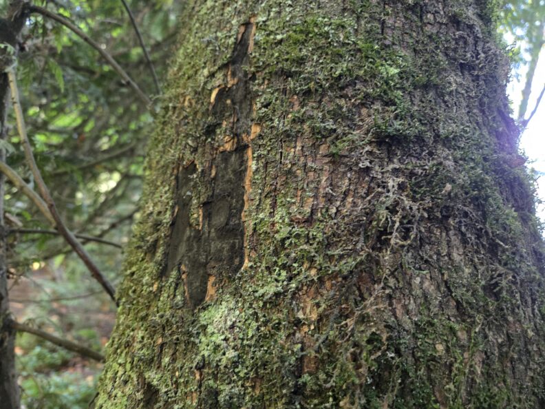
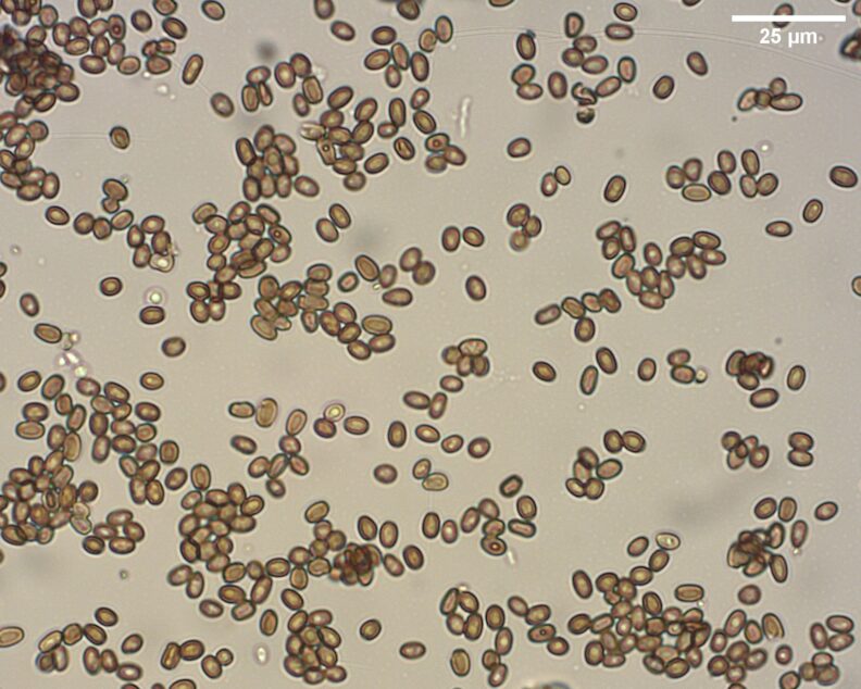
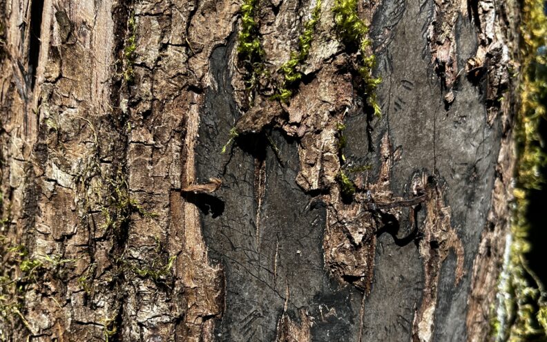
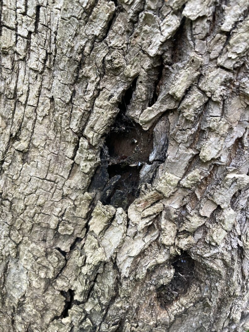
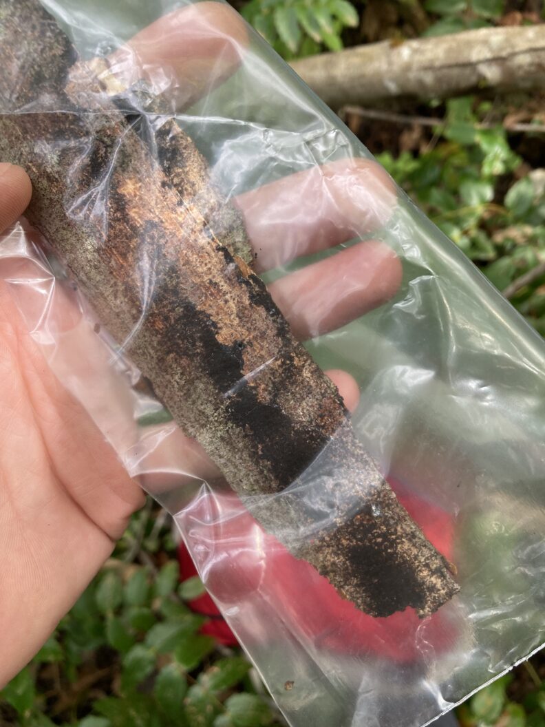
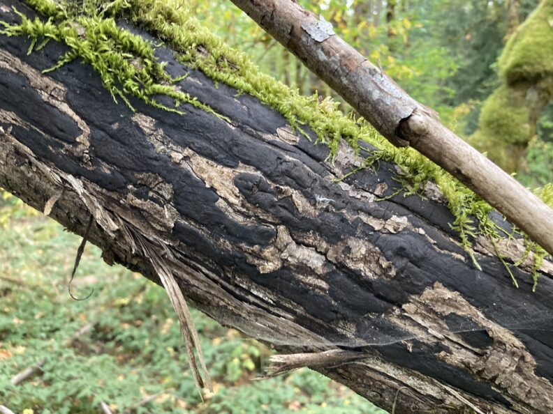
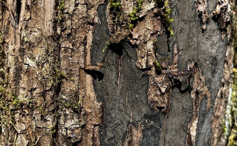
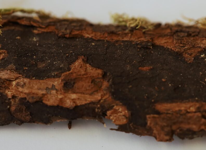
1. Spores and Lesions
Within the bark of the infected branch or stem, large amounts of black “soot-like” spores begin to accumulate. Eventually, the buildup of these spores causes the outer layer of the bark to shed, revealing black lesions covered in millions of spores. Occasionally, white spores are seen, although the cause of this different color is unknown. Eventually, all the spores will be blown away by wind, leaving behind a weathered lesion.


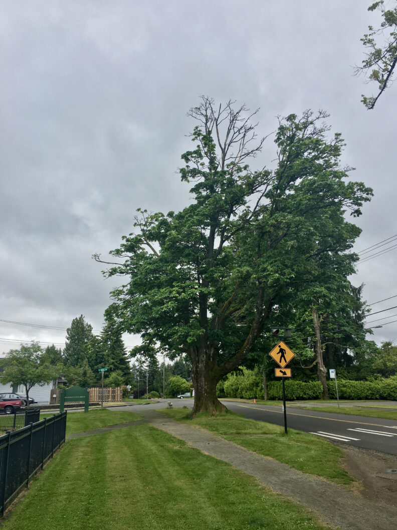
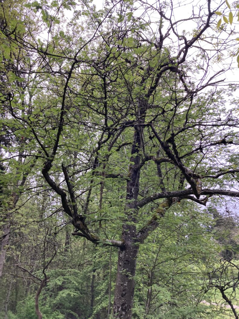
2. Tree Decline
Often, trees with SBD will experience symptoms such as foliar dieback, foliar stunting, and other signs of overall tree decline. These symptoms are fairly non-specific and are not always indicative of SBD! Abiotic stressors, pests, and other diseases may cause similar symptoms.
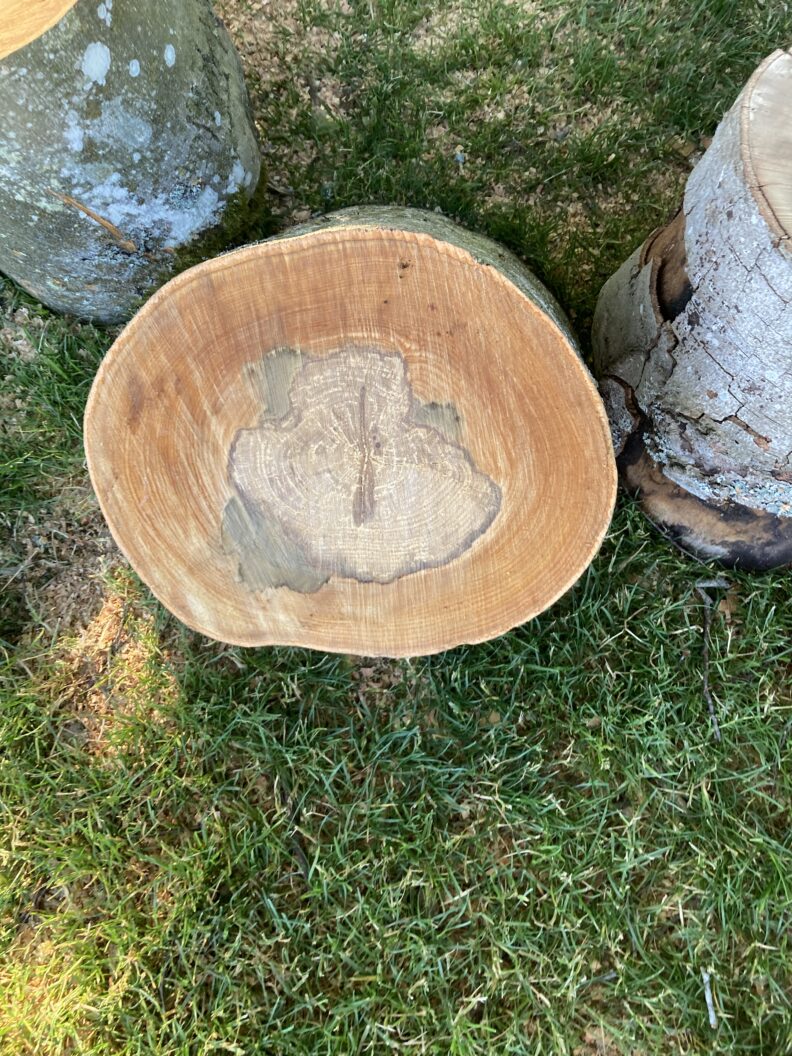
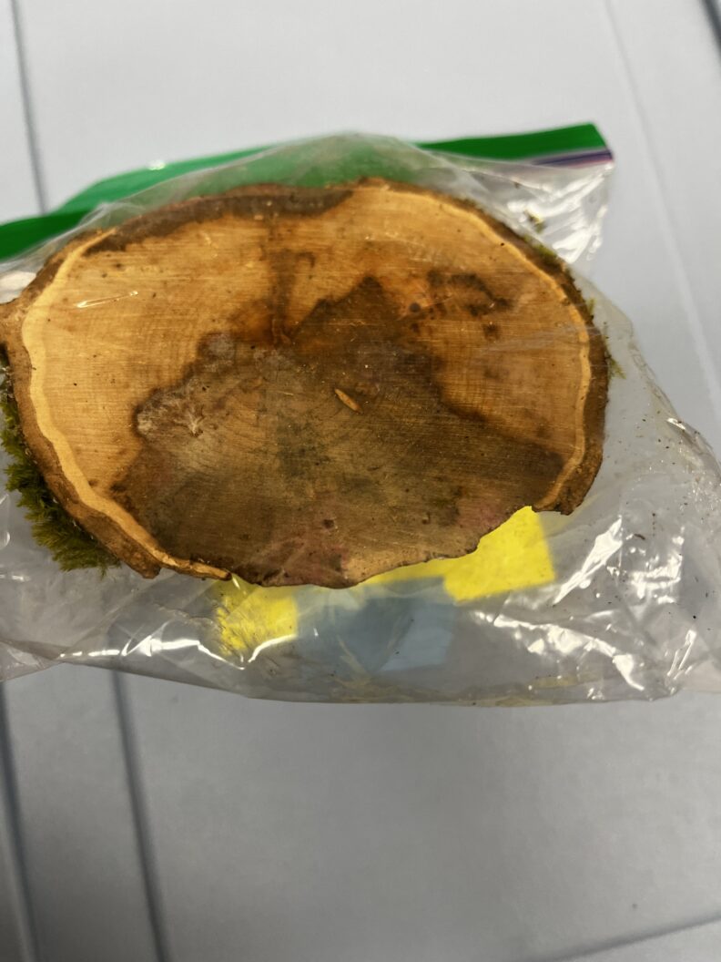
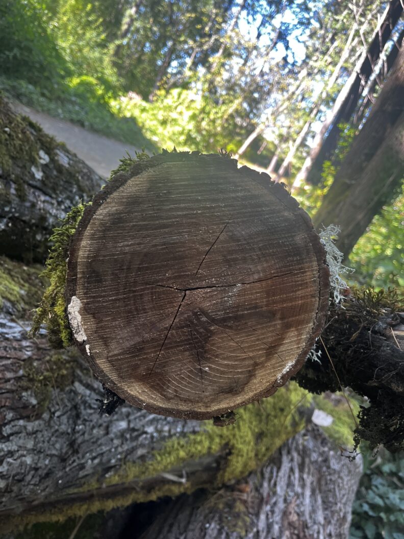
3. Internal Staining
In freshly cut branches and stems, a brown stain with a green leading edge is often seen through the cross section of the sample. As the cross-section begins to dry out, the stain loses its green coloration.

Interested in Submitting a Sample?
1. Identify trees with signs and symptoms of SBD
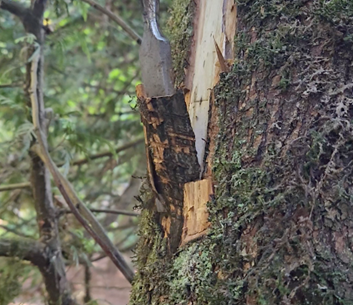
Trees with SBD typically show crown die-back and overall decline. The most easily identifiable sign of disease is the presence of black cankers on the inner layer of bark.
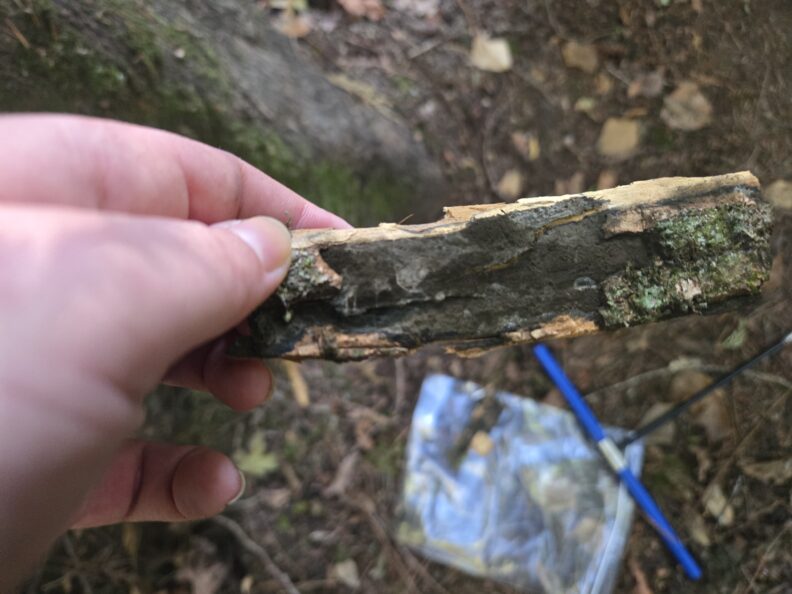
2. Collect the sample
Sampling with a chisel or flathead screwdriver is usually the best way to get a good sample. Take approximately a 3 inch x 3 inch sample of the wood along the edge of the lesio. Try to sample the woody tissue, and include a sample of the out bark, if possible.
Take photos of all symptoms visible on the tree of interest. Additionally, record the name of the tree (scientific or common name), tree condition (alive or dead), and GPS coordinates, if possible.
3. Submit your sample
Samples can be sent to:
ATTN: Taylor McNees/ Joey Hulbert
WSU Puyallup Research and Extension Center
2606 West Pioneer
Puyallup, WA, 98371-4998 USA
Please send us an email (taylor.mcnees@wsu.edu) for any questions, and to let us know to expect some samples from you.
Close Look-a-likes
Species of Biscogniauxia can easily be mistaken for the SBD fungus! These species are closely-related to Cryptostroma corticale and often form similar black lesions on stumps, fallen logs, and sometimes living trees. However, there are a few key differences between lesions cause by these species.
Differentiating Cryptostroma and Biscogniauxia
Biscogniauxia species
- Lesions are crust-like and tend to form on top of the bark
- Small spore like structures are often seen on the lesion
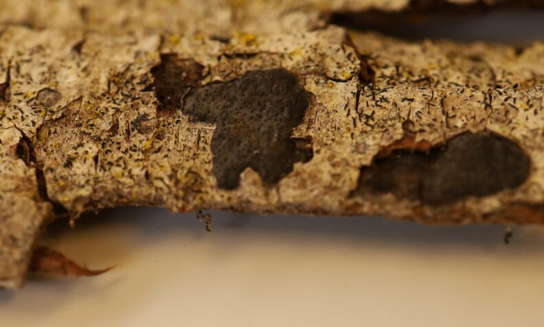
Cryptostroma corticale
- Lesion forms on innermost layer of the bark (not crust-like)
- No visible pore-like structures on the lesion. Lesion often covered in black “soot-like” spores
Seeking Collaborators
In search of samples from across the USA
We are looking to obtain samples of C. corticale from across the United States for use in a population genomics study to better understand the genetic diversity and movement of the fungus throughout the country.
Please contact Taylor McNees if you’re interested in collaborating! Taylor is a PhD Student in Plant Pathology at WSU studying the population genetics of Cryptostroma corticale.
Our Goal:
To collect samples of Cryptostroma corticale, the causal agent of Sooty Bark Disease, from across the United States to use in a population genomics study.
Questions we hope to answer with this research:
1. Where is Crytsostroma corticale native to?
It is currently hypothesized that C. corticale is native to Great Lakes region of North America due to first being described in London, Ontario. We predict that we will be able to use genomic data to better deduce the native region of C. corticale within the United States.
2. How has Cryptostroma corticale moved throughout the continental United States?
Currently, C. coticale has been identified in 3 states – California, Michigan, and Washington. We hypothesize that by collecting nationwide samples we can gain a better understanding of both how C. corticale is distributed and how it has moved across the country.
3. Is sexual reproduction occurring within the species?
C. corticale belongs to a group of fungi known to produce both sexual and asexual spores. However, sexual spores of C. corticale have not been identified! We predict that population genomics data will provide insight into whether sexual reproduction and genetic recombination of this fungus is occurring.
Human Health: Maple Bark Disease
Chronic inhalation of spores has been linked to the development of Maple Bark Disease (MPD), a type of hypersensitivity pneumonitis, in humans. For more information on the risks of MPD, please visit the following links:
- Washington State Department of Health – Maple Bark Disease
- Sooty Bark Disease in BC: A cause of a rare form of hypersensitivity pneumonitis
Additional Resources
Relevant webpages, news articles, and diagnostic guides
- Barlett Tree Experts – Sooty Bark Disease
- Sooty Bark Disease Diagnostic Guide
- Pacific Northwest Pest Management Handbook – Maple (Acer spp.) – Sooty Bark Disease
Technical literature
- Kelnarová, I., Černý, K., Zahradník, D., Koukol, O. (2017). Widespread latent infection of Cryptostroma corticale in asymptomatic Acer pseudoplatanus as a risk for urban plantations. Forest Pathology 47:e12344. https://doi.org/10.1111/efp.12344
- Brooks, R. K., Omdal, D., Brown, S., Marshall, C. J., Hulbert, J. M., Elliott, M., & Chastagner, G. (2023). Cryptostroma corticale, the causal agent of sooty bark disease of maple, appears widespread in western Washington State, USA. Forest Pathology, 53(6), e12835. https://doi.org/10.1111/efp.12835
- Ogris, N., Brglez, A., & Piškur, B. (2021). Drought stress Can Induce the Pathogenicity of Cryptostroma corticale, the Causal Agent of Sooty Bark Disease of Sycamore Maple. Forests, 12(3), Article 3. https://doi.org/10.3390/f12030377
- Braun, M., Klingelhöfer, D., & Groneberg, D. A. (2021). Sooty bark disease of maples: The risk for hypersensitivity pneumonitis by fungal spores not only for woodman. Journal of Occupational Medicine and Toxicology, 16(1), 2. https://doi.org/10.1186/s12995-021-00292-5
- Tanney, J. B., Feau, N., Shamoun, S. F., Kope, H. H., Dicaire, A., Drugmand, B., Walker, J., Burlakoti, P., & Joshi, V. (2024). Cryptostroma corticale (Ellis & Everh.) P. H. Greg. & S. Waller causing sooty bark disease in British Columbia, Canada. Canadian Journal of Plant Pathology, 46(6), 596–610. https://doi.org/10.1080/07060661.2024.2369324
Contact Us
Please reach out to Taylor McNees (taylor.mcnees@wsu.edu) with any questions or for more information!
Samples can be sent to:
ATTN: Taylor McNees/ Joey Hulbert
WSU Puyallup Research and Extension Center
2606 West Pioneer
Puyallup, WA, 98371-4998 USA
Please email us to let us know to expect a sample from you.
Page last updated: October 22nd, 2025.